Contrast Enhanced Ultrasound (CEUS)

Radiation-Free. Real-Time. Remarkably Sensitive.
CEUS is a cutting-edge imaging modality that uses microbubble contrast agents to highlight vascularity in real time—without radiation. It offers unmatched sensitivity to blood flow, especially in lesions or regions that grayscale ultrasound might miss.
Advantages of CEUS

Radiation-free technique
Real-time imaging
Robust enhancement patterns
Cutting-edge technology
Used across liver, kidney, male & female pelvic organs
Bedside imaging capability
Safe for patients with renal failure
Highly sensitive to microbubble contrast
Low incidence of contrast reactions
Repeatable for follow-up
Lower wait times for imaging
Ideal for claustrophobic or critically ill patients
Precision in Motion,
Clarity in Vision.

Clinical Leadership

Research Champion
Dr. Ahmed El Kaffas
View
Clinical Champion
Dr. Aman Khurana
View
View
Senior Faculty Contributor
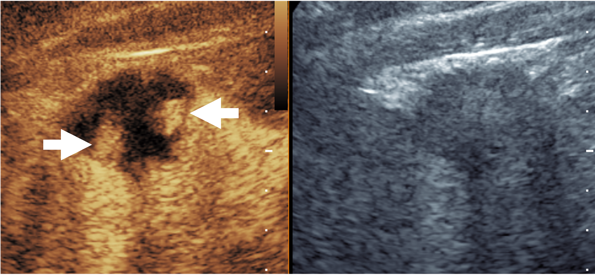
Figure 1 — Contrast Enhanced US (CEUS) and paired grayscale US images showing robust contrast enhancement within the nodular peripheral areas of viable tumor (arrows) in a treated hepatic cavity just 30 seconds after the microbubble injection.
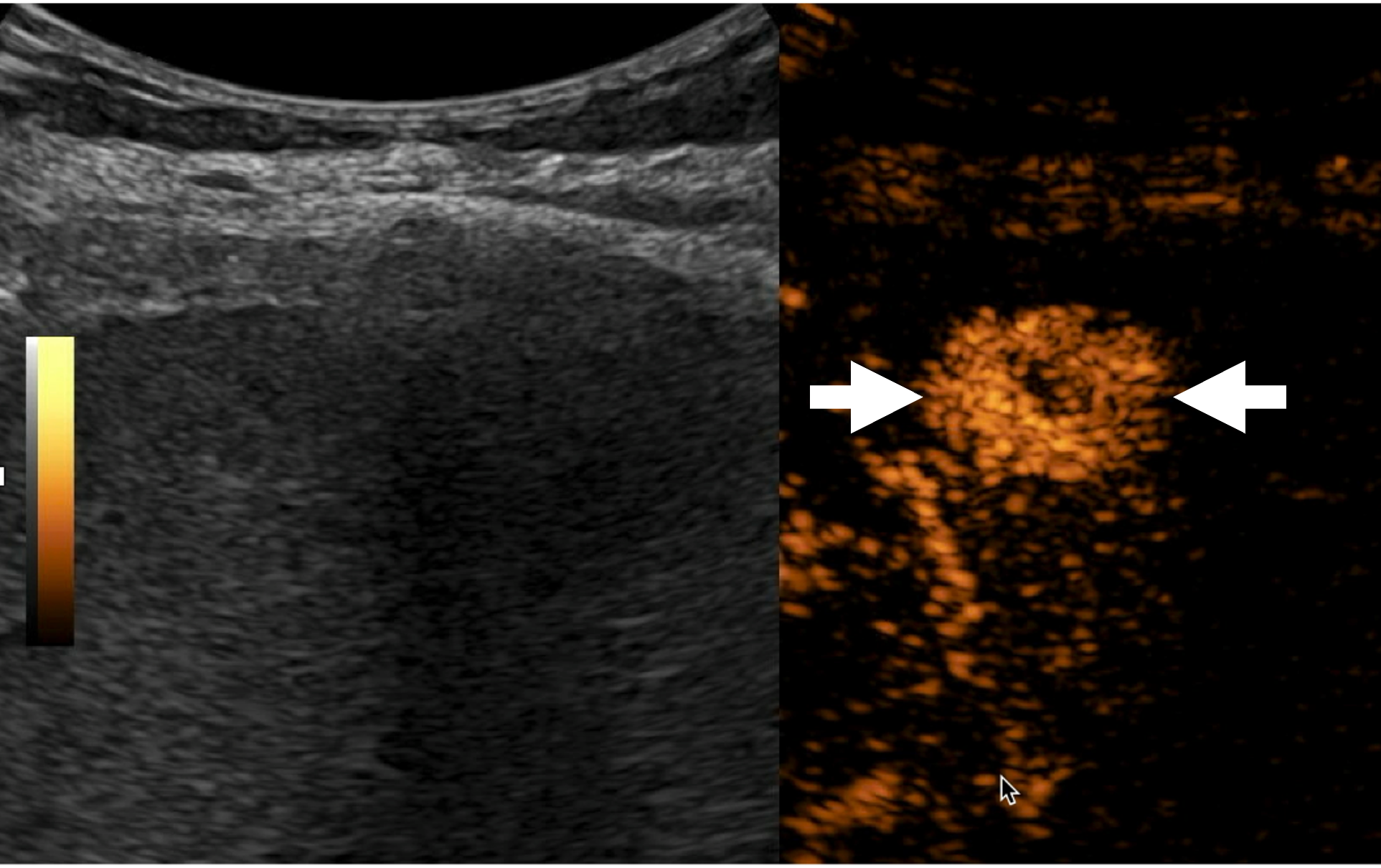
Figure 2 — Paired grayscale US and CEUS images showing remarkable sensitivity of this modality to its’ contrast agent where a benign liver lesion enhances significantly only 14 seconds after the microbubble contrast injection (arrows). Please note that lesion is nearly impossible to see on conventional grayscale US.
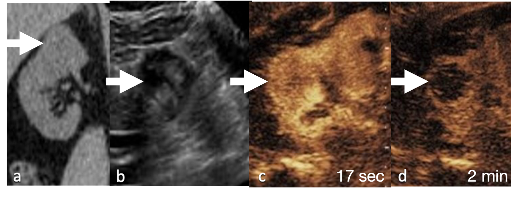
Figure 3 — Non contrast CT shows an indeterminate lesion arising from the superior pole right kidney (arrow). Grayscale ultrasound (b) shows a solid mass with mixed echogenicity (arrow). CEUS shows enhancement (arrow) in the early arterial phase at 17 seconds (c) post microbubble injection with washout (arrow) at 2minutes (d), consistent with renal cell carcinoma, later pathologically proven as papillary renal cell carcinoma.
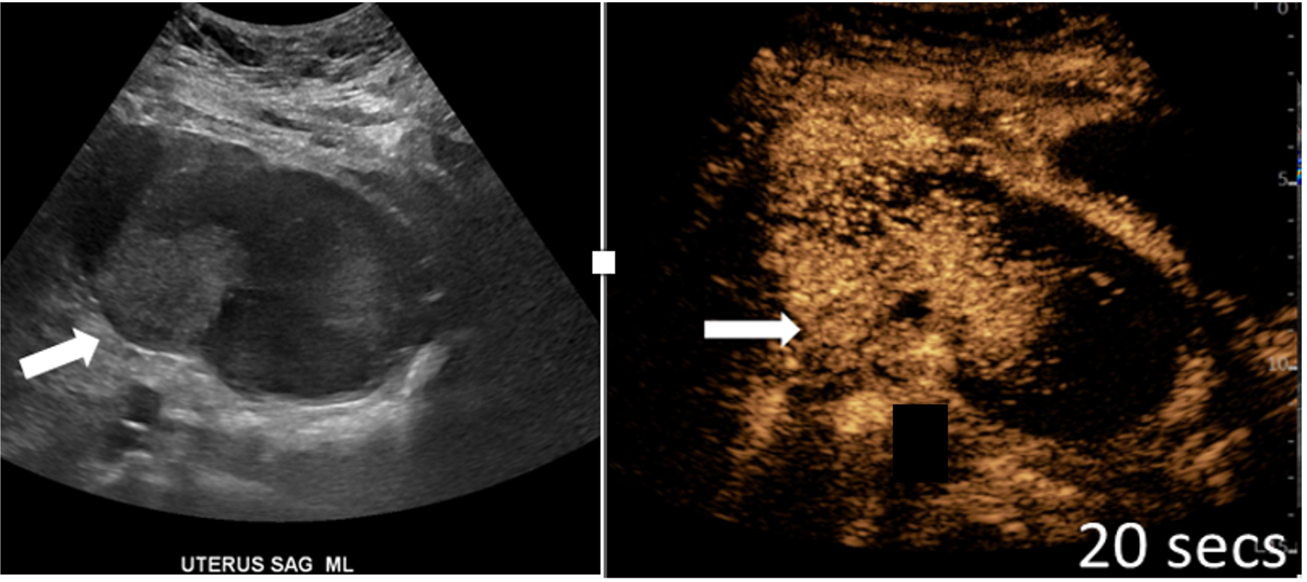
Figure 4 — Paired grayscale US and CEUS images of the uterus insagittal view demonstrating a hyperechoic endometrial mass with early arterial hyperenhancement at 20 seconds post microbubble contrast injection with invasion of the myometrium(arrow).
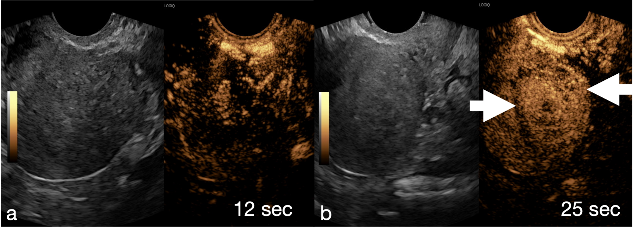
Figure 5 — Paired grayscale US and CEUS images of the uterus insagittal view at 12 (a) and 25 seconds (b) after microbubble contrast injection demonstrating an ill-defined hyperechoic lesion with synchronous and rapid enhancement of the entire lesion (arrows) consistent with typical imaging findings of a focal uterine adenomyoma.

Performance

Enhanced Imaging Clarity
Provides superior visualization of organs and blood flow.
Non-Invasive and Safe
No radiation exposure; safe for repeated use.
Real-Time Results
Instant feedback during examinations.
Patient-Friendly
Ideal for patients with kidney failure, fear of closed spaces, and contrast allergies.
Accuracy
05 / 05
Non-Invasiveness
05 / 05
Real-Time Feedback
05 / 05
Patient Comfort
04 / 05
Reliability
05 / 05

Technology

Microbubble Contrast Agents
Enhance ultrasound signals by reflecting sound waves for clearer images.
Real-Time Data Processing
Enables immediate visualization of organs and blood flow.
Advanced Signal Enhancement
Delivers high-definition images for more accurate diagnoses.
Alternative Imaging Option
Suitable for patients sensitive to traditional contrast dyes.

Specifications

Maximum Imaging Depth
15 cm
Resolution
Sub-millimeter
Scanning Time
15–30 minutes
System Compatibility
Most ultrasound systems

Applications

Liver and Kidney Imaging
Detects tumors, cysts, and vascular abnormalities.
Cardiac Imaging
Assesses heart function and blood flow.
Oncology Monitoring
Tracks cancer progression and treatment response.
Pediatric Imaging
Safe for children due to the absence of radiation.