MVP-ASL: Advanced MRI of Cerebral Blood Flow

Unlocking the future of brain health through MRI technology.
MVP-ASL (MicroVascular Pulsatility and Velocity-Selective Arterial Spin Labeling) is a breakthrough technique that captures something conventional imaging simply can’t: the subtle rhythmic pulse of blood coursing through the brain’s tiniest vessels.

By pairing velocity-selective arterial spin labeling with retrospective cardiac gating, MVP-ASL synchronizes every image frame to the patient’s heartbeat. The result is a dynamic, beat-by-beat map of cerebral blood flow and microvascular pulsatility—an early, quantitative marker of cerebrovascular health.
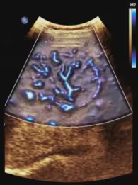
Animation illustrating how MVP-ASL visualizes the brain's blood flow pulsing with each heartbeat.
Higher blood flow is shown in red, while lower flow is depicted in blue.
Transitions between high and low flow occur dynamically throughout the cardiac cycle.

Technology

dynamic cerebral blood flow
Visualize patterns of cerebral blood flow across the heartbeat
Microvascular Pulsatility Mapping
Detailed blood flow pulsatility information across brain regions
3D Volumetric imaging
High-resolution, 3-D images of blood flow pulsatility
Contrast-free imaging
Safe for adult and pediatric patients

Transformative Potential

Novel Biomarker for Brain Health
Cerebral blood flow pulsatility measured with MVP may offer insight into dementia and other neurodegenerative conditions
Predicting Cognitive Decline
MVP-ASL holds promise for identifying individuals at risk of future cognitive decline
Non-Invasive
Safe and comfortable for all patients, with no need for dyes or harmful radiation

Specifications

Maximum Imaging Depth
Unlimited
Resolution
2-3 millimeter
Scanning Time
7 minutes
System Compatibility
1.5T and 3T MRI

Key Attributes

Novelty
05 / 05
Non-Invasiveness
05 / 05
Speed of Analysis
04 / 05
Patient Comfort
04 / 05
Diagnostic Potential
05 / 05

Clinical Leadership

Research Champion
Dr. Conan Chen
View
Clinical Champion
Dr. Divya S. Bolar
View
View
Senior Faculty Contributors
Dr. Thomas T. Liu
View
Dr. Eric C. Wong
View

The MVP-ASL Approach

Beat-by-beat mapping of the brain’s microvascular pulse
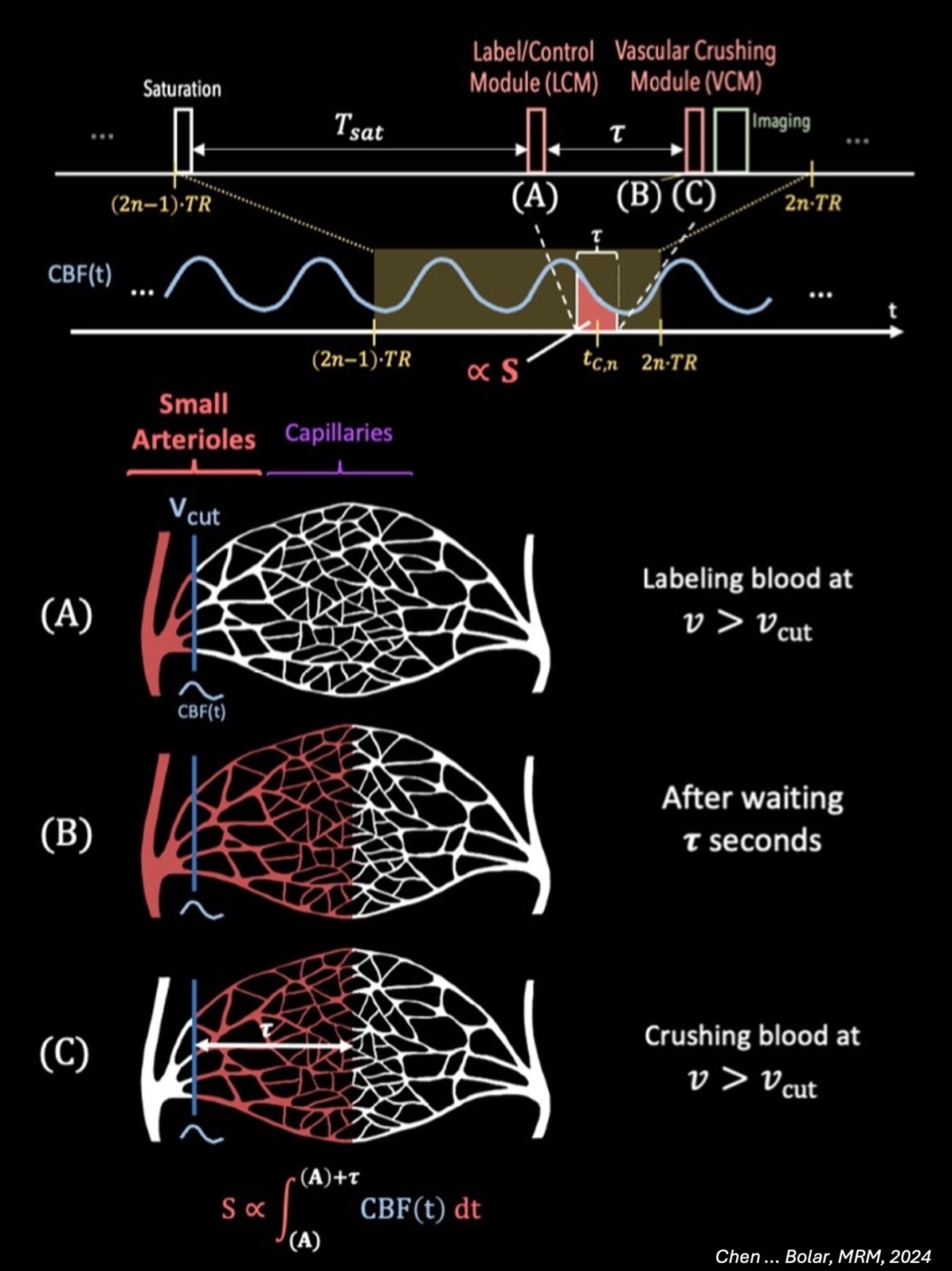
This figure illustrates how MVP-ASL—MicroVascular Pulsatility with Velocity-Selective Arterial Spin Labeling—combines velocity-selective labeling with retrospective cardiac gating so that MRI can capture a complete perfusion map on every heartbeat.

By synchronizing each image frame to the cardiac cycle, the sequence quantifies MicroVascular Pulsatility (MVP), a beat-level waveform of blood flow in arterioles and capillaries that standard MRI cannot resolve.

This beat-by-beat view enables earlier detection of microvascular dysfunction, delivers dynamic quantitative biomarkers for monitoring stroke, dementia, and small-vessel therapies, and provides a potential early indicator of neurodegenerative risk—linking subtle vascular changes to conditions such as Alzheimer’s disease.

Core Parameters

Velocity-selective labeling: $v_c = 2$ cm/s cutoff threshold

Temporal Resolution: $\approx 30$ ms per cardiac phase

Spatial Resolution: 2–3 mm isotropic voxels

Signal Equations

MVP-ASL Signal:

$$S(t) = S_0 \left(1 - 2\alpha \cdot e^{-TI/T_1} \cdot CBF(t)\right)$$

Variables:
$S_0$ = equilibrium magnetization
$\alpha$ = labeling efficiency
$TI$ = post-labeling delay
$T_1$ = longitudinal relaxation time
$CBF(t)$ = time-varying cerebral blood flow

Quantification Methods

Pulsatility Index (PI):

$$PI = \frac{CBF_{max} - CBF_{min}}{CBF_{mean}}$$

Temporal Features:
• Time-to-peak (TTP)
• Full-width at half-maximum (FWHM)
• Mean transit time (MTT)

Validation Metrics

Signal Quality:
SNR $> 15$ in gray matter
CV $< 5\%$ temporal stability

Reliability:
ICC $> 0.85$ spatial reproducibility
$> 90\%$ test-retest reliability